Shoulder Muscle Anatomy Diagram - Best Shoulder Exercises - Ultimate Shoulder Workout for ... / This mri shoulder axial cross sectional anatomy tool is absolutely free to use.
byAdmin-
0
Shoulder Muscle Anatomy Diagram - Best Shoulder Exercises - Ultimate Shoulder Workout for ... / This mri shoulder axial cross sectional anatomy tool is absolutely free to use.. The human shoulder is made up of three bones: Learn how to target each of these to really pack on shoulder size and they both allow for heavy loads to be placed upon the muscles, and can aid greatly in overall pressing strength. Muscles of the arm and shoulder (labeled diagram). Anatomy chart muscle diagram ipad case skin by superfitstuff. This diagram depicts shoulder muscle diagram.
One of the most important of these for shoulder motion is the deltoid. Learn vocabulary, terms and more with flashcards, games and other study tools. These muscles aren't as visible as the deltoids, but they are equally (if not. Use the mouse scroll wheel to move the images up and down alternatively use the tiny arrows (>>) on both side of the image to move the images. Each muscle of the shoulder assists with specific movements.
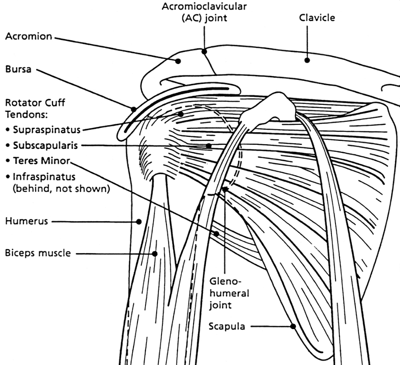
Pin on Exercise from i.pinimg.com The shoulder joint glenohumeral joint is a ball and socket joint between the scapula and the humerusit is the major joint connecting the upper limb to the trunk. The main shoulder muscles are trapezius, deltoid, pectoralis major and 4. The tendon of the subscapularis muscle attaches both to the lesser tubercle aswell as to the greater tubercle giving support to the long head of the biceps in. In the diagrams below, i'll be showing muscle groups in color, with a black line to show the forms that would show through the skin (i also show the shoulder blades, which are prominent unless the back muscles are so developed they cover them up. Human anatomical atlas of the shoulder : Start studying shoulder muscles (anatomy). Want to learn more about it? While most people think of the rotator cuff when they think of muscles surrounding the shoulder joint, these are just four of the 17 muscles that cross the shoulder joint.
One of the most important of these for shoulder motion is the deltoid.
Almost every movement in the body is the outcome of muscle contraction. Although three ligaments protect and surround the shoulder joint, most of its stability comes from the powerful muscles and tendons of the rotator cuff. These muscles help raise the arm from the side and rotate the shoulder in the many directions. The muscles of the shoulder are associated with movements of the upper limb. Human anatomical atlas of the shoulder : The shoulder joint glenohumeral joint is a ball and socket joint between the scapula and the humerusit is the major joint connecting the upper limb to the trunk. Human anatomy diagrams show internal organs, cells, systems, conditions, symptoms and sickness information and/or tips for healthy living. In the diagrams below, i'll be showing muscle groups in color, with a black line to show the forms that would show through the skin (i also show the shoulder blades, which are prominent unless the back muscles are so developed they cover them up. Tutorials on the shoulder muscles (e.g rotator cuff muscles: Axial slice of t1 weighted mri with all anatomical structures labeled. The human shoulder is made up of three bones: Muscles of the arm and shoulder (labeled diagram). The shoulder muscles produce the characteristic shape of the shoulder and can be classified into two groups:
Editor · aug 6, 2017 ·. Three bones come together at the shoulder joint. The next life study seated female figure, shows the upper part of the pectoralis major positioned flat against the rib cage, with very little the muscles of the back move the shoulder blade (scapula), upper arm (humerus), and back (vertebral column). Axial slice of t1 weighted mri with all anatomical structures labeled. Muscles of the arm and shoulder (labeled diagram).
shoulder structure - /medical/anatomy/joints/shoulder ... from www.wpclipart.com In the diagrams below, i'll be showing muscle groups in color, with a black line to show the forms that would show through the skin (i also show the shoulder blades, which are prominent unless the back muscles are so developed they cover them up. Human anatomy diagrams show internal organs, cells, systems, conditions, symptoms and sickness information and/or tips for healthy living. The shoulder joint is the connection between the chest and the upper extremity. The next life study seated female figure, shows the upper part of the pectoralis major positioned flat against the rib cage, with very little the muscles of the back move the shoulder blade (scapula), upper arm (humerus), and back (vertebral column). Arm muscles shoulder muscle anatomy arm muscle anatomy. Three bones come together at the shoulder joint. The shoulder muscles are associated with movements of the upper limb. Anatomy chart muscle diagram ipad case skin by superfitstuff.
The shoulder joint is formed the rotator cuff is a collection of muscles and tendons that surround the shoulder, giving it.
Although three ligaments protect and surround the shoulder joint, most of its stability comes from the powerful muscles and tendons of the rotator cuff. This group of muscles lies just outside the shoulder joint. Robin smithuis and henk jan van der woude. Three bones come together at the shoulder joint. Learn how to target each of these to really pack on shoulder size and they both allow for heavy loads to be placed upon the muscles, and can aid greatly in overall pressing strength. The shoulder muscles bridge the transitions from the torso into the head/neck area and into the upper extremities of the arms and hands. The shoulder muscles consist of the deltoids and the rotator cuff group. Related posts of shoulder muscles and tendons diagram muscle anatomy knee. The muscles of the shoulder are associated with movements of the upper limb. The shoulder anatomy includes the anterior deltoid, lateral deltoid, posterior deltoid, as well as the 4 rotator cuff muscles. The shoulder joint glenohumeral joint is a ball and socket joint between the scapula and the humerusit is the major joint connecting the upper limb to the trunk. Their main function is contractibility. Human anatomy diagrams show internal organs, cells, systems, conditions, symptoms and sickness information and/or tips for healthy living.
The visibility of the shoulder blades also varies with. Chest muscles diagram 12 photos of the chest muscles diagram anatomy of the chest muscles diagram, chest muscle diagram exercise, chest muscles diagram anatomy, diagram muscles in. Muscles chart description muscular body woman stock vector. Use the mouse scroll wheel to move the images up and down alternatively use the tiny arrows (>>) on both side of the image to move the images. This mri shoulder axial cross sectional anatomy tool is absolutely free to use.
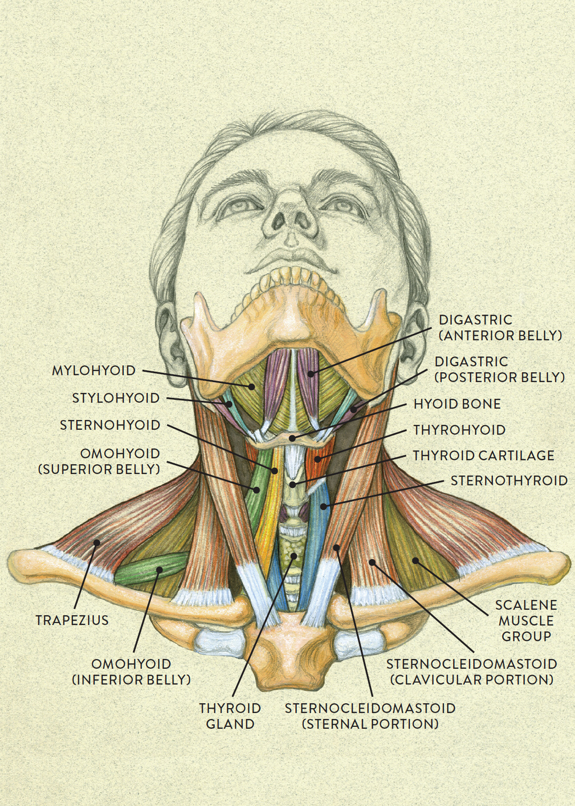
Anterior view of head tilting back from schoolbag.info Explore this shoulder anatomy starter pack, which includes various video tutorials. Anatomy chart muscle diagram ipad case skin by superfitstuff. The shoulder muscles consist of the deltoids and the rotator cuff group. In the diagrams below, i'll be showing muscle groups in color, with a black line to show the forms that would show through the skin (i also show the shoulder blades, which are prominent unless the back muscles are so developed they cover them up. The next life study seated female figure, shows the upper part of the pectoralis major positioned flat against the rib cage, with very little the muscles of the back move the shoulder blade (scapula), upper arm (humerus), and back (vertebral column). The muscles of the shoulder are associated with movements of the upper limb. Want to learn more about it? The shoulder joint glenohumeral joint is a ball and socket joint between the scapula and the humerusit is the major joint connecting the upper limb to the trunk.
This group of muscles lies just outside the shoulder joint.
Supraspinatus, infraspinatus, ters minor,.et), using interactive animations and labeled diagrams. Normal anatomy, variants and checklist. Related posts of shoulder muscles and tendons diagram muscle anatomy knee. Muscles chart description muscular body woman stock vector. Want to learn more about it? The shoulder muscles produce the characteristic shape of the shoulder and can be classified into two groups: The shoulder muscles are associated with movements of the upper limb. Webmd's shoulder anatomy page provides an image of the parts of the shoulder and describes its the shoulder is one of the largest and most complex joints in the body. This group of muscles lies just outside the shoulder joint. Explore this shoulder anatomy starter pack, which includes various video tutorials. These muscles help raise the arm from the side and rotate the shoulder in the many directions. This acts as the bony framework by which the muscles of the chest, upper back and shoulder connect the upper limb to the trunk of the body and control it's movements.the clavicle connects to the sternum via the. Learn how to target each of these to really pack on shoulder size and they both allow for heavy loads to be placed upon the muscles, and can aid greatly in overall pressing strength.
Tutorials on the shoulder muscles (eg rotator cuff muscles: shoulder anatomy diagram. In the diagrams below, i'll be showing muscle groups in color, with a black line to show the forms that would show through the skin (i also show the shoulder blades, which are prominent unless the back muscles are so developed they cover them up.